Figure 9
OPEN and CLOSED state of SPIKE SARS-COV-2: relationship with some integrin binding. A biological molecular approach to better understand the coagulant effect
Luisetto M*, Khaled Edbey, Mashori GR, Yesvi AR and Latyschev OY
Published: 02 July, 2021 | Volume 5 - Issue 1 | Pages: 049-056
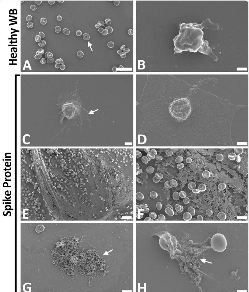
Figure 9:
9: Scanning electron- micrographs of healthy control whole- blood (WB), with and without spike protein. A and B) Healthy WB smears, with arrows indicating normal erythrocyte- ultrastructure. C to H) Healthy WB exposed to spike- protein (1 ng.mL-1 final concentration), with C and D) indicating the activated -platelets (arrow), E and F) showing the spontaneously formed fibrin- network and G and H) the anomalous deposits that is amyloid in nature (arrows) (Scale bars: E: 20μm; A: 10μm; F and G: 5μm; H: 2μm; C: 1μm; B and D: 500nm). Form article SARS-CoV-2 spike S1 subunit induces hyper-coagulability SARS-CoV-2 spike protein S1 induces fibrin (ogen) resistant to the fibrinolysis: Implications for microclot formation in COVID-19 Lize M. Grobbelaar, Chantelle Venter, Mare Vlok, Malebogo Ngoepe, Gert Jacobus Laubscher, Petrus Johannes Lourens, Janami Steenkamp Douglas B. Kell, Etheresia Pretorius Preprint. Wool GD, Miller JL. The Impact of COVID-19 Disease on Platelets and Coagulation Pathobiology 2021; 88: 15–27.
Read Full Article HTML DOI: 10.29328/journal.abb.1001028 Cite this Article Read Full Article PDF
More Images
Similar Articles
-
OPEN and CLOSED state of SPIKE SARS-COV-2: relationship with some integrin binding. A biological molecular approach to better understand the coagulant effectLuisetto M*,Khaled Edbey,Mashori GR,Yesvi AR,Latyschev OY. OPEN and CLOSED state of SPIKE SARS-COV-2: relationship with some integrin binding. A biological molecular approach to better understand the coagulant effect . . 2021 doi: 10.29328/journal.abb.1001028; 5: 049-056
Recently Viewed
-
COVID-19 Vaccines Development: Challenges and Future PerspectiveSwapan Kumar Chowdhury,Niranjan Kumar Mridha,Abdul Ashik Khan,Nabajyoti Baildya,Manab Mandal*,Narendra Nath Ghosh*. COVID-19 Vaccines Development: Challenges and Future Perspective. Int J Clin Virol. 2025: doi: 10.29328/journal.ijcv.1001064; 9: 010-020
-
The Synergistic Effect of Combined Linagliptin and Metformin Improves Hepatic Function in High-fat Diet/Streptozotocin-induced Diabetic RatsFolasade Omobolanle Ajao*,Ifedolapo Opeyemi Adeyeye,Noheem Olaoluwa Kalejaiye,Sodik Olasunkami Mukaila,Olalekan Samson Agboola,Marcus Olaoye Iyedupe. The Synergistic Effect of Combined Linagliptin and Metformin Improves Hepatic Function in High-fat Diet/Streptozotocin-induced Diabetic Rats. Ann Clin Gastroenterol Hepatol. 2025: doi: 10.29328/journal.acgh.1001050; 9: 004-012
-
Schizoaffective Disorder in an Individual with Mowat-Wilson Syndrome (MWS)Yadwinder Chuhan, Nimrit Bath, Muhammad Ayub*. Schizoaffective Disorder in an Individual with Mowat-Wilson Syndrome (MWS). Arch Psychiatr Ment Health. 2024: doi: 10.29328/journal.apmh.1001050; 8: 008-011
-
Clinical Severity of Sickle Cell Anaemia in Children in the Gambia: A Cross-Sectional StudyLamin Makalo,Samuel A Adegoke,Stephen J Allen,Bankole P Kuti,Kalipha Kassama,Sheikh Joof,Aboulie Camara,Mamadou Lamin Kijera,Egbuna O Obidike. Clinical Severity of Sickle Cell Anaemia in Children in the Gambia: A Cross-Sectional Study. J Hematol Clin Res. 2025: doi: 10.29328/journal.jhcr.1001033; 9: 001-006
-
Prospective evaluation of a computerized algorithm for Vitamin K antagonist drug dose calculationMaarten J Beinema*,Jacobus RBJ Brouwers,Henk Adriaansen,Frank GA Jansman. Prospective evaluation of a computerized algorithm for Vitamin K antagonist drug dose calculation. J Hematol Clin Res. 2023: doi: 10.29328/journal.jhcr.1001020; 7: 001-005
Most Viewed
-
Feasibility study of magnetic sensing for detecting single-neuron action potentialsDenis Tonini,Kai Wu,Renata Saha,Jian-Ping Wang*. Feasibility study of magnetic sensing for detecting single-neuron action potentials. Ann Biomed Sci Eng. 2022 doi: 10.29328/journal.abse.1001018; 6: 019-029
-
Evaluation of In vitro and Ex vivo Models for Studying the Effectiveness of Vaginal Drug Systems in Controlling Microbe Infections: A Systematic ReviewMohammad Hossein Karami*, Majid Abdouss*, Mandana Karami. Evaluation of In vitro and Ex vivo Models for Studying the Effectiveness of Vaginal Drug Systems in Controlling Microbe Infections: A Systematic Review. Clin J Obstet Gynecol. 2023 doi: 10.29328/journal.cjog.1001151; 6: 201-215
-
Prospective Coronavirus Liver Effects: Available KnowledgeAvishek Mandal*. Prospective Coronavirus Liver Effects: Available Knowledge. Ann Clin Gastroenterol Hepatol. 2023 doi: 10.29328/journal.acgh.1001039; 7: 001-010
-
Causal Link between Human Blood Metabolites and Asthma: An Investigation Using Mendelian RandomizationYong-Qing Zhu, Xiao-Yan Meng, Jing-Hua Yang*. Causal Link between Human Blood Metabolites and Asthma: An Investigation Using Mendelian Randomization. Arch Asthma Allergy Immunol. 2023 doi: 10.29328/journal.aaai.1001032; 7: 012-022
-
An algorithm to safely manage oral food challenge in an office-based setting for children with multiple food allergiesNathalie Cottel,Aïcha Dieme,Véronique Orcel,Yannick Chantran,Mélisande Bourgoin-Heck,Jocelyne Just. An algorithm to safely manage oral food challenge in an office-based setting for children with multiple food allergies. Arch Asthma Allergy Immunol. 2021 doi: 10.29328/journal.aaai.1001027; 5: 030-037

HSPI: We're glad you're here. Please click "create a new Query" if you are a new visitor to our website and need further information from us.
If you are already a member of our network and need to keep track of any developments regarding a question you have already submitted, click "take me to my Query."


